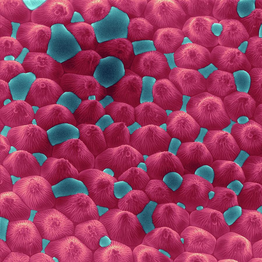
Rose Petal Upper Surface, SEM - Stock Image - F017/4073 - Science Photo Library
Papillae on the upper surface of a rose flower petal (Rosa sp), coloured scanning electron micrograph (SEM). Papillae are projections from epidermal cells and in the rose they are conical in shape. DENNIS KUNKEL MICROSCOPY/SCIENCE PHOTO LIBRARY

Focused ion beam scanning electron microscopic image of the rose petal

A) SEM image of structures found on the surface of rose petals. The

Petal upper surface hi-res stock photography and images - Alamy

Rose petal, SEM - Stock Image - C016/2667 - Science Photo Library

Evaluation of minerals, toxic elements and bioactive compounds in rose petals (Rosa spp.) using chemometric tools and artificial neural networks - ScienceDirect

4 SEM images of the red rose petal surface showing the micropapillae

Petal upper surface hi-res stock photography and images - Alamy

Focused ion beam scanning electron microscopic image of the rose petal

Petal upper surface hi-res stock photography and images - Alamy

Rose Petal Upper Surface #5 Photograph by Dennis Kunkel Microscopy/science Photo Library - Fine Art America

Rose petal surface. Each surface cell is 20 microns across! via @wellcomeimages
Rose Petal With Water Droplets Photograph by Dennis Kunkel Microscopy/science Photo Library
Rose Petal With Water Droplets by Dennis Kunkel Microscopy/science Photo Library

Rose Petal Upper Surface, SEM - Stock Image - F017/4072 - Science Photo Library