Left): Porcine ventricle sample, epicardium side up, mounted to the

The three myocardial slices shown at the top were harvested from the
Biomimetics, Free Full-Text

Quantifying the microstructural and biomechanical changes in the porcine ventricles during growth and remodelling - ScienceDirect
The mesh of the left and right ventricle, showing epicardium and RV and
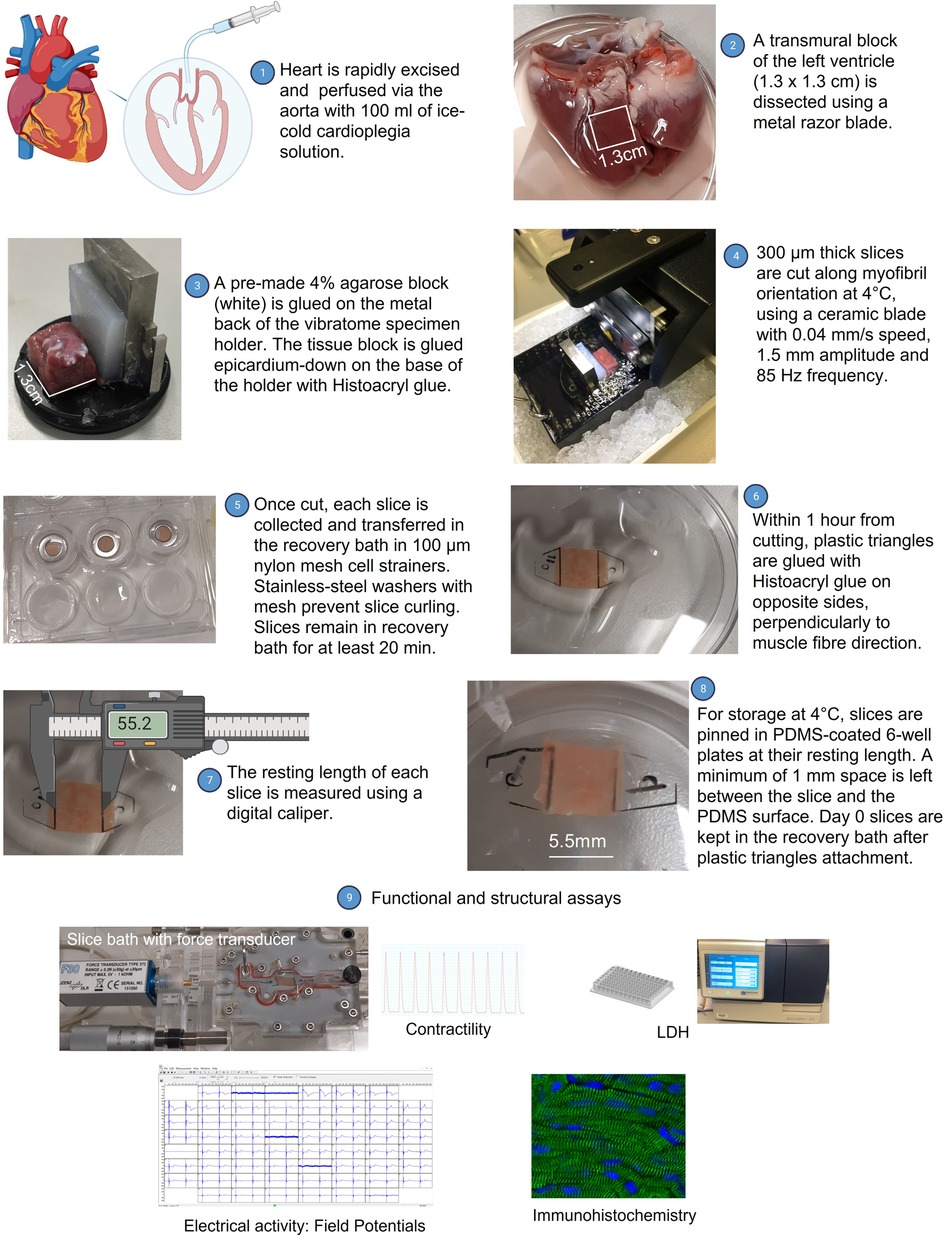
Frontiers A novel method to extend viability and functionality of living heart slices

Cardiac Stromal Cell Patch Integrated with Engineered Microvessels Improves Recovery from Myocardial Infarction in Rats and Pigs

PDF) Anatomy of the pig heart: Comparisons with normal human cardiac structure

Section levels of the left ventricle.

Anatomical and molecular mapping of the left and right ventricular His–Purkinje conduction networks - Journal of Molecular and Cellular Cardiology

Heart - Wikipedia
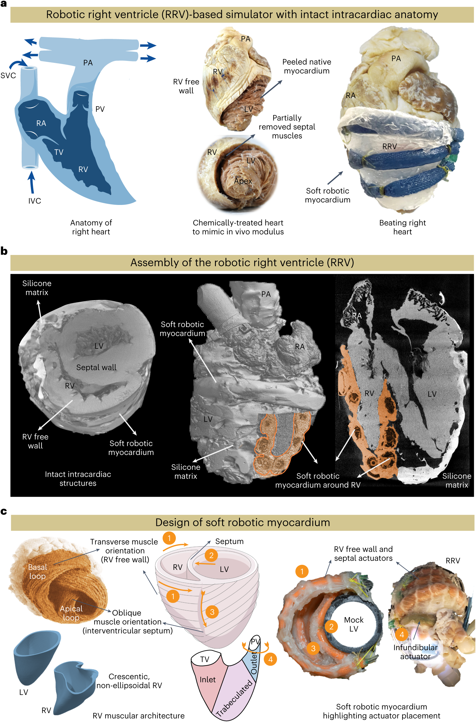
Robotic right ventricle is a biohybrid platform that simulates right ventricular function in (patho)physiological conditions and intervention

Morphology, distribution, and variability of the epicardiac neural ganglionated subplexuses in the human heart - Pauza - 2000 - The Anatomical Record - Wiley Online Library

The Left and Right Ventricles

Preparation of a small mammalian (rat) left ventricular tissue block.

Comparative Cardiac Anatomy